Department of Biocybernetic system


Mission and Research Areas of the Biocybernetics Systems Department:
The aims of biocybernetics department are to investigate mechanisms of interaction of the biological and technological systems, modeling and control with interdisciplinary approach, which integrates biology, cybernetics, engineering, and AI. The department’s aim is creating of such knowledge, which will promote medicine, biotechnologies and neuro- engineering in Georgia and outside.
Biomedical Investigations
Prostate cancer
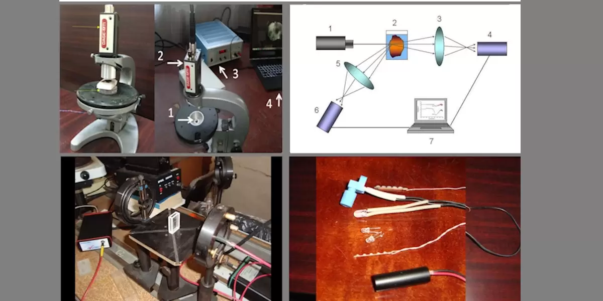
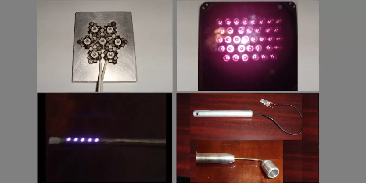
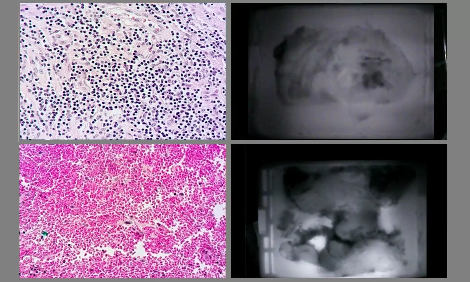
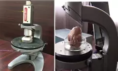
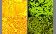
Conventional techniques (PSA, MRI, TRUS) frequently fail to provide an accurate cancer diagnosis at an early stage. Based on the results of our research, it was determined that the optical density of malignant tissue is higher than that of healthy tissue. These results made it feasible to develop the novel prostate cancer visualization technique. This led to the development and fabrication of the pilot (model) equipment for prostate cancer visualization-diagnostics. The device uses infrared light to illuminate the prostate from the inside. We obtain infrared images of the prostate using a device with a CCD camera and our proprietary software. The gadget can accurately identify cancers that are 2-3 mm in size. It may be suggested for targeted biopsies.
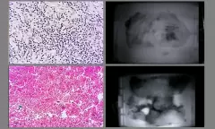
The aggressiveness of prostate cancer is assessed using the established approach. The results obtained are entirely consistent with the Glisson score sum approach.
Kidney cancer
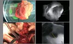
In modern kidney cancer treatment, partial nephrectomy is commonly used. This procedure involves removing only the tumor and a small margin of surrounding healthy tissue. The goal is to ensure that the entire tumor is removed, leaving no cancer cells behind. Traditionally, this is verified during surgery using the intraoperative frozen section (FS) method, where tissue samples are analyzed under a microscope. FS typically takes 20–25 minutes because blood vessels must be clamped during the procedure, increasing the risk of warm ischemia and postoperative complications.
We have developed a new method based on infrared imaging that clearly distinguishes between cancerous and healthy tissue. This technique takes only 5–6 minutes to perform, offering a significant advantage over FS.
Bio electromagnetism studies
The second area of our research, known as bio electromagnetism studies, focuses on the effects of various electromagnetic frequencies on cells, animals, and people.
The primary emphasis of research on bio electromagnetics is:
Our research focuses on studying the effects of high frequency electromagnetic fields on neurons, tiny birds, insects, and humans.
The investigations include:
1.Digital modeling and simulation of how various biological objects are exposed to electromagnetic fields.
- Developing methods to determine the specific absorption rate (SAR).
- Modeling human exposure based on simulations, including scenarios such as being inside a car, using a mobile phone, and experiencing thermal effects.
- Studying how antennas interact with parts of the human body—like the hand or fingers—and how this affects the antenna’s radiation characteristics, which is crucial for understanding real-world exposure.
- Numerical dosimetry, which involves analyzing SAR levels in small biological objects exposed to radiofrequency electromagnetic fields. This helps us understand the impact of 5G and other wireless technologies on different biological species.
- Investigating how radio waves and microwaves affect neuronal activity and information processing in neurons.